


“Our research combines cutting-edge genetics with heritage science, using X-ray CT to reveal barley’s internal structure in unprecedented detail. This breakthrough not only advances crop science for climate resilience but also opens new possibilities for studying ancient plant remains and preserving botanical collections.”
Professor Lisa-Marie Shillito, Durham University
Context and challenge
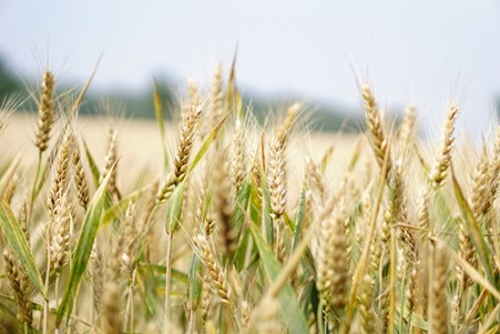
Understanding how crops respond to environmental stress is a central challenge for both modern agriculture and the study of past farming systems. Barley, one of the earliest domesticated cereals, provides a vital link between ancient agricultural practices and contemporary crop improvement.
However, traditional analytical methods have limited researchers’ ability to observe internal plant structures at cellular scale without damaging rare or fragile specimens. By addressing this challenge with X-ray Computed Tomography (XR-CT), researchers have integrated insights from past agricultural practices with modern crop science, supporting UKRI’s commitment to building sustainable and resilient food systems.
Investment in action
The North East Material Culture Analytical Suite (NEMCAS) team at Durham University mobilising AHRC Capability for Collections (CapCo) investment alongside current RICHeS investment to provide access to XR-CT facilities and specialist expertise.
This investment enabled NEMCAS to support an interdisciplinary UKRI-BBSRC funded research project led by Dr Peter Etchells, bringing together plant genetics, archaeology and heritage science. Without sustained infrastructure investment, the advanced imaging capabilities required for this research would not have been accessible to the project.
Innovative methods
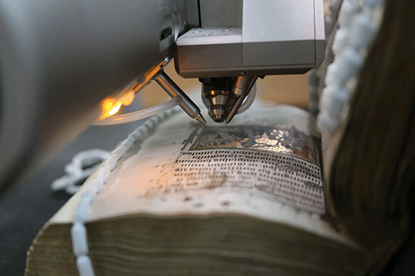
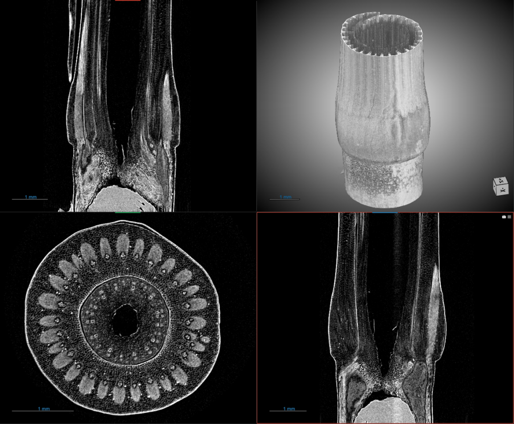
Using XR-CT, a non-destructive imaging technique, the team captured high-resolution, three-dimensional images of barley stem architecture at micrometer resolution. XR-CT allows intact barley stems to be scanned and analysed, revealing internal cell structures in exceptional detail. By integrating archaeological expertise and advanced imaging, the team achieved a level of structural resolution previously unattainable in living plants, while establishing methods directly applicable to ancient plant remains.
Unlocking outcomes
This project advances knowledge and demonstrating how shared research infrastructure can contribute to global challenges in food security, climate resilience, and sustainable agriculture.
The use of this non-destructive technique enables fragile seeds or charred archaeological specimens to be examined while preserving rare physical specimens. This has enabled detailed comparison between barley varieties, linking differences in cell architecture to the function of specific genes and understanding traits such as stress tolerance and grain quality. These datasets inform preservation, enhance understanding of ancient agricultural practices and offer a deep-time perspective on crop resilience.
With research ongoing, the work is expected to generate further benefits including expanded interdisciplinary collaboration, technology sharing and potential commercial opportunities.