


Cover image © Trustees of the British Museum
The RICHeS team recently visited the British Museum where they met with Dr Dan O’Flynn, Head of Multiscale X-ray Imaging Centre and the project lead for the RICHeS’ funded facilities project Multiscale Heritage X-ray Imaging Centre. Beneath the public galleries, the RICHeS team explored how this project is set to enhance the museum’s existing heritage science capabilities through the development of a cutting-edge micro-CT laboratory, opening the door to exciting new collaborations.
A hidden hub of heritage science capabilities
Below the galleries of the British Museum lies a remarkable suite of heritage science facilities. These laboratories are located within the World Conservation and Exhibitions Centre that opened in 2014, and home to the Museum’s Scientific Research department. Here, world-leading researchers work to care for millions of artefacts in the collection, while also providing analytical services to external partners.
The facilities include a wide range of advance imaging and analytical tools including a photogrammetry rig for 3D surface imaging, and a Reflectance Transformation Imaging (RTI) dome for capturing miniscule surface details and variations. Additionally, the department has laboratories for Scanning Electron Microscopy (SEM), X-ray diffraction and X-Ray Fluorescence (XRF), techniques that offer unique insights into the composition, construction and sometimes hidden features of historic objects.
The facilities also feature a newly upgraded mass spectrometer, supported by the Arts and Humanities Research Council Capability for Collections (CapCo) Fund. Mass spectrometry is a powerful analytical technique that helps scientists understand the molecular makeup of materials. This CapCo investment provided a high-resolution GC-QTOF-MS (Gas Chromatography – Quadrupole Time-of-Flight – Mass Spectrometry) system that enables precise identification of organic compounds and molecular structures that are essential for materials research in heritage science. Research arising from the use of the new instrument has already been reported helping to provide museum scientists and conservators with key information to accurately identify Pistacia resin and preserve objects containing it.
Powering discovery with X-ray imaging
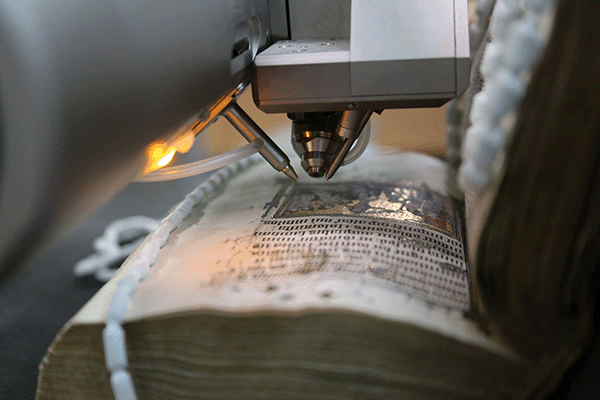
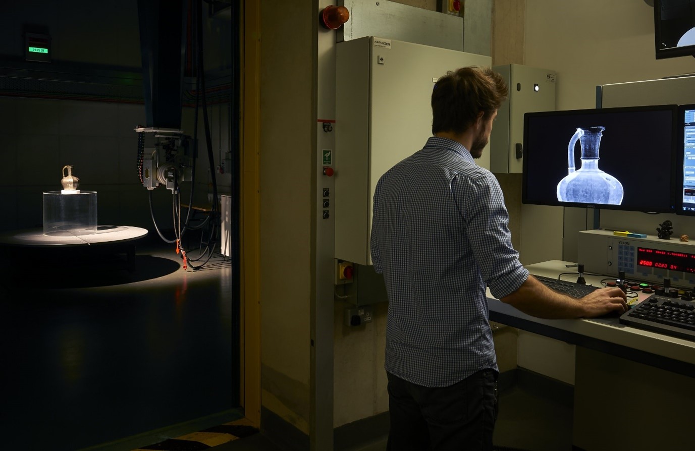
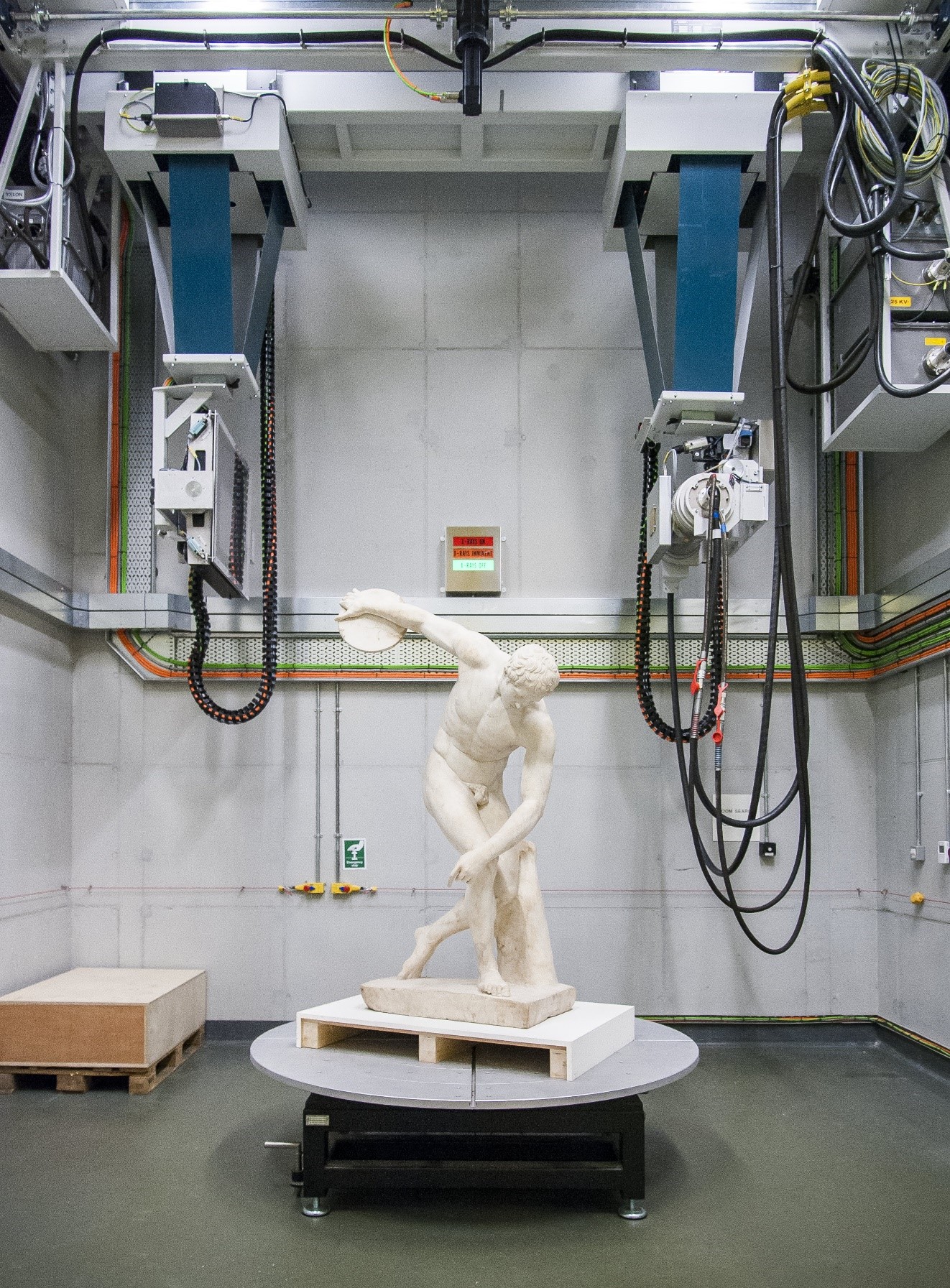
The RICHeS team visit concluded with a tour of the museums large scale X-ray imaging facility. X-rays have played a key role in heritage science since their discovery in the 1890s, and the British Museum’s on-site facility continues this tradition on a much more advanced scale. The facility is spacious enough to scan large sculptures and powerful enough to image dense materials like metals.
With concrete shielding walls, a lead-lined door and robust safety procedures, the facility handles equipment capable of generating radiation up to around three times the voltage of a hospital Computed Tomography (CT) scan. Its capabilities have already supported groundbreaking research. For example, the powerful scans have revealed unseen imagery in the Galloway Hoard for National Museums Scotland.
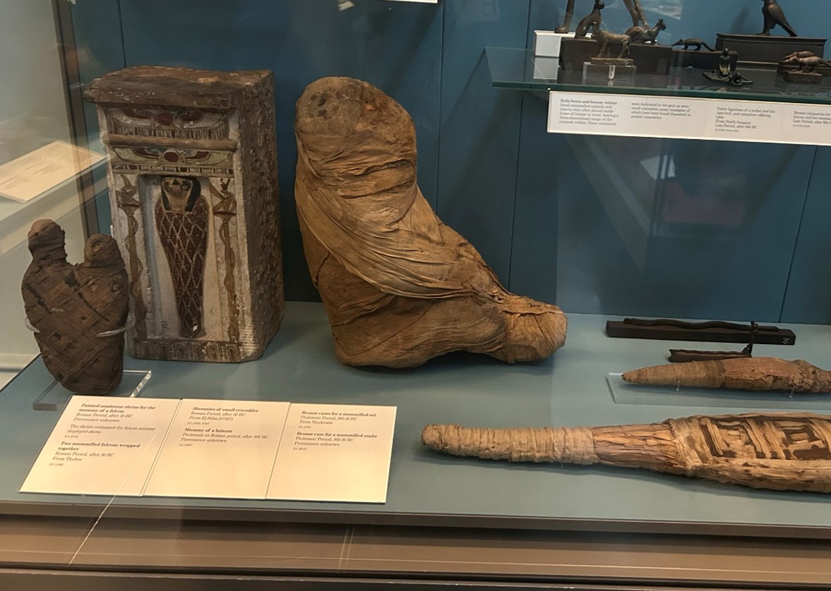
While the current facility already includes some CT functionality, the RICHeS investment will elevate this with the introduction of a dedicated micro-CT laboratory. This will allow for high-resolution 3D imaging of smaller objects. These innovative CT scans are central to several ongoing research projects at the Museum, including Divine creatures: animal mummies in ancient Egypt , Rethinking the Lewis chess pieces in the North Sea and Arctic world and Making cuneiform tablets. Among the examples shown during this visit was a mummified baboon that was first x-rayed in 1899. The findings from X-ray imaging offer remarkable insights into ancient practices and provide compelling content for public engagement.
Opening up access and collaboration
The new RICHeS funded micro-CT facility, integrated with existing labs, will benefit from bespoke data infrastructure to handle the high-resolution scan outputs. As a key partner in the Heritage Science Data Service (HSDS) consortium, the British Museum is also contributing to the broader digital infrastructure that will support heritage science research across the UK.
A new staff role, funded through the RICHeS programme will help expand access to the imaging facilities at the British Museum while also supporting collaborations with other institutions across the county. The RICHeS IHQ team are excited to see how these developments will foster new research, deepen collaborative discoveries and strengthen the UK’s distributed heritage science infrastructure.
Dr Dan O’Flynn shares:
“X-ray imaging gives us fantastic potential for uncovering the stories held inside heritage collections, and for connecting in new ways with public audiences. Thanks to generous support from the AHRC, we are very excited to be establishing the Multiscale X-ray Imaging Centre at the British Museum, which will enable us to see more than ever under the surface, and to open up these possibilities to the heritage science community“